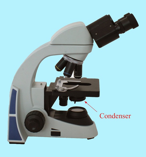
Microscope condensers consist of one or more lenses located directly under the microscope stage. By focusing the light rays from the scattered microscope source, the lenses produce a tidy cone or beam of light that shines through the stage aperture and specimen.
The operation of most microscopes is based on two factors. The first is that the light is hitting the specimen appropriately. Second, the correct amount of light reaches the objective lens. The specimen must be properly illuminated by a light source. The light must refract in a systematic manner through and around the specimen. It is important to organize the light so that it can reflect into the objective lens so that the information is absorbed efficiently?
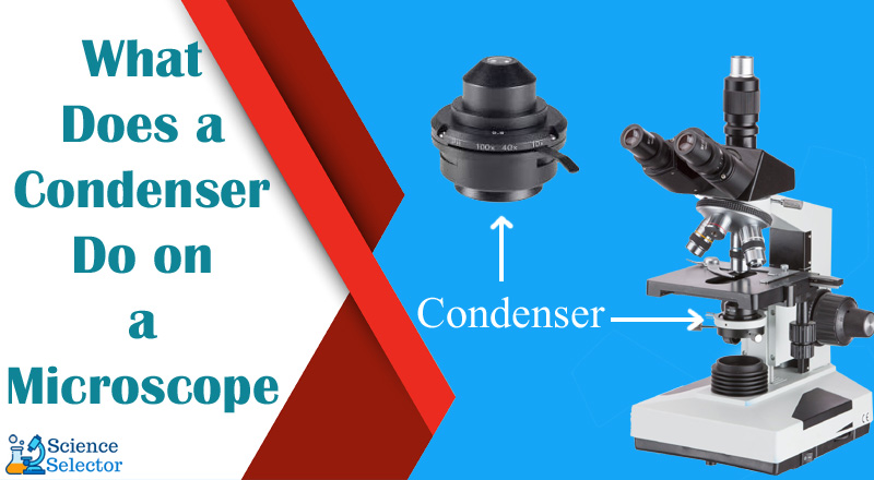
The light passes through several stages on its way from the light source to the specimen. A halogen or LED illumination is a straightforward light source. To achieve the best results, we need to focus the light on the sample, which is much smaller than the size of the light source. The condenser is used for this. We explore what does a condenser do on a microscope in this article. Read on to learn more.
The Condenser
The condenser’s job is to modify light’s direction and angles of reflection, not to enhance it in any way. The condenser regulates how much light from the illuminator can travel through the aperture, hence regulating the light’s intensity. It also regulates the contrast, which is crucial.
The contrast between distinct, drab-colored objects in the visual field is most significant in darkfield microscopy, not their appearance per se. They’re used to pluck out images that wouldn’t show up if the equipment was just used to blast the slide with as much light as the eyes above it could handle, leaving the spectator to hope for the best.
Optical Elements Of A Microscope
Objective Lenses
These are all the microscope’s major lenses. Magnification ranges from 4x to 100x, with three to five lenses in most cases. These objective lenses can be looking backward or forwards.
Stage
The stage is where you put the specimen or item that you want to look at. A mechanical stage is required when working at greater magnifications and requiring tiny motions of the sample or slide.
Aperture
Aperture is the opening that allows light to pass through so that it can reach the stage.
Illuminator
The light source for the microscope is the Illuminator, which is located at the bottom of the instrument. Halogen bulbs are commonly used in light microscopes, and the control for these bulbs is located at the bottom of the microscope.
Condenser Iris Diaphragm
The amount of light that reaches the specimen is controlled by the condenser iris diaphragm. The iris diaphragm of the condenser can be found above and below the stage, respectively. An Abbe condenser and an iris diaphragm are common features of high-end microscopes. The condenser and diaphragm of the microscope work together to regulate the amount and focus of light hitting the sample.
Condenser Focus Knob
The focus knob on the condenser moves it up and down, which focuses the light on the specimen.
Stage Clips
Using stage clips is important if the viewer is manually repositioning the slide to look at different regions of the sample
Eyepiece or Ocular
The eyepiece, also known as the ocular, is located at the top of the microscope and is used to see the sample under examination. The normal eyepiece has a magnification of 10x, but there are alternative eyepieces available with magnifications ranging from 5x all the way up to 30x or more!
Eyepiece Tube
The eyepiece tube is responsible for keeping the objective lens in place while using a microscope. A diopter adjustment ring is usually included with a binocular microscope (an optical microscope having two eyepieces that can alleviate eye strain). This piece of gear will compensate for any discrepancies in your vision that you may have.
Using a microscope while wearing glasses, for example, is difficult. A diopter will not be included in a single eye (monocular) microscope. Binocular microscopes, because no two persons have the same Interpupillary distance, allow for this customization.
Nosepiece
This is the area of the microscope where the actual lenses are located. The rotating turret holds the objective lenses, making it considerably simpler to select the one you need. The most common goals are 4x, 10x, 40x, and 100x. There are numerous objectives with various magnifications of power available.
Coarse and Fine Focus knobs
The Coarse Focus knob brings the sample close to being focused while the Fine Focus brings it fully into focus. Using the Fine focus knob, you may then fine-tune and sharpen the image’s sharpness.
Types of Condensers
There are a variety of condensers available to adjust for aberrations or flaws that are visible to the spectator. The condenser, as we now know, aids in focusing the light onto the specimen in a more organized and uniform manner. To avoid getting too technical, this focused process might be challenging.
Because light rays don’t always travel in a straight line, engineers and scientists had to get creative with condenser design to accommodate for these flaws. The Abbe condenser, Aplanatic condensers, and specialized condensers are the three main types of condensers.
Abbe Condenser
The prototype Abbe Condenser was originally named after Ernst Abbe, who developed it in the 1800s. This is the same condenser we discussed in the last section.
These are the standard condensers that come with entry-level microscopes because they’re the cheapest option.
Spherical and chromatic aberrations are not corrected by these sorts of condensers, although the higher the price, the greater the numerical aperture can be. Take a look at this post if you’re not familiar with numerical aperture.
Aplanatic and Achromatic Condensers
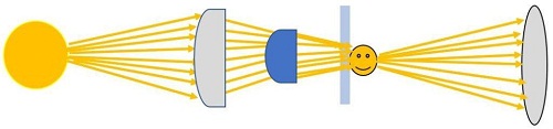
Light focus faults can be addressed with condensers of this type. The light that enters and leaves the condenser lenses should look like this:
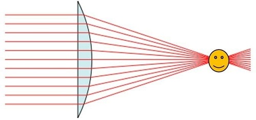
After passing through the second lenses fairly parallelly, the light beams condense and emerge on the smiling face. This actually occurs in reality.
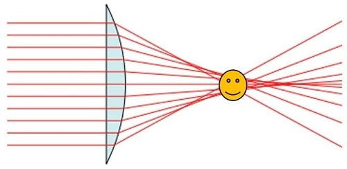
There’s no light coming through here! An aberration is a technical term used by optical engineers to describe something like this. No lens will ever be perfect because of flaws in the glass substance or the way the lens is fashioned. To compensate for light source distortions, you would utilize one of these advanced condensers to ensure flawless light enters your sample.
A spherical aberration is what we have here. Condensers, then, are devices that help to reduce mistakes in light propagation. Chromatic aberrations are a different sort of aberration.
For this, there are a few more variables to consider. A slight deviation from the ideal path is seen in spherical aberration, where all light appears to travel in an “off-path” fashion. On the other hand, chromatic aberrations cause every light ray, regardless of wavelength, to deviate from its ideal course.
Due to light diffracting in and out of the lenses, this happens. Chromatic aberration-solving condensers fix every wavelength’s light path, which is an improvement over spherical aberration-solving condensers.
Specialized Condensers
Finally, we have a rather broad category! Microscopes can be used for all kinds of specialized tasks. The use of different kinds of light sources such as LED, OLED, or halogen lights as the light source should be the same whether you are looking at transparent specimens, light or dark specimens, and so on.
If you are probing dark materials, you will have to correct for how the light interacts with the sample if you are using condensers like darkfield or phase contrast. Condensers are being used in all sorts of ways, you just have to research the one that fits what you are trying to do.
How Does a Condenser Work?
Observe the light that comes out of the light bulb. What do you notice? It can go in whatever way you want it to! If we’re trying to direct the light upwards through the stage apertures, this won’t work.
As much of this light as possible must be focused on the specimen of interest. To begin, let’s put together a simple design that includes a light source and a specimen to be studied.
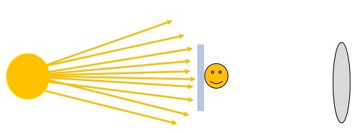
From what we can make out, light isn’t getting to the specimen very well (the happy face on the glass slide). They’re coming at it from all directions and angles. The light will be disorganized when it goes through the microscope slide and into the lens on the other side, resulting in a hazy, pixelated image.
The condenser plays an important role here. Between the light source and the specimen will be a condenser that “converges” the random angles. It organizes all of the scattered light into a single focused beam of light.
As a result of the focusing, a more even distribution of light strikes the sample. Thus, when we look at a specimen with our eyes after it has passed through the objective lens, it appears more clear and vivid because of the light that has exited the specimen. A condenser is a pair of lenses, although a single lens may be included in a less expensive microscope set. Light is collected by a bigger lens, which also serves as a light source, while a second, smaller lens sharpens the light for a closer focus.
When it comes to controlling the condenser, there are usually two controls to consider. One knob controls how close or far the condenser is to the specimen stage. By doing this, you can fine-tune the image’s resolution as you see fit. The diaphragm, on the other hand, regulates the amount of light that reaches the sample. The Diaphragm of a Microscope: An Introduction to Diaphragms provides additional information about diaphragms. What exactly is it, and how can you put it to use in your life?
Here’s a quick rundown of how a microscope condenser works mechanically:
- A light source’s rays travel randomly.
- The condenser lens is exposed to some light that hits the lens.
- As the light passes through the condenser lens, convergent or parallel beams are created.
- An organized beam of light is emitted from this point.
- An “organized” cone of light is transmitted to the objective lens through the specimen, where it is refracted.
Adjusting Light Intensity For A Microscope Condenser
When the light intensity is regulated correctly, a microscope condenser will work to its full potential. Lower magnification levels typically necessitate less illumination.
The condenser’s performance can be affected by the sort of light you utilize in your microscope. LED lights, on the other hand, will have a significantly higher luminance than fluorescent light. Changing the microscope’s light intensity setting is a simple process.
FAQ
What is the difference between the condenser and the iris diaphragm?
The use of field diaphragms can help ensure proper microscope alignment, but they are not typically included in more affordable models. When it comes to controlling contrast, the aperture diaphragm (sometimes referred to as an iris diaphragm) lives in the condenser, which is located beneath the microscope stage and in line with the instruments’ objectives.
What is the condenser on a microscope quizlet?
Condenser. The specimen is illuminated by a small non-magnifying lens situated beneath the stage. The light output may be adjusted by raising and lowering the condenser. The finest place to stand is towards the stage’s lower edge.
Is the condenser above or below the iris diaphragm?
It’s placed below the stage, above the condenser. An iris diaphragm and an Abbe condenser are standard features in most high-end microscopes. Their combined effect regulates the amount of light directed at the specimen while also controlling its focus.
What is the function of the condenser in a brightfield microscope quizlet?
It receives light from the source at the bottom and concentrates it into a cone that illuminates the sample. The knob to the left of the condenser adjusts the quantity of light that hits the specimen.
When should microscope lenses be cleaned?
You should probably have it cleaned by a professional if the spot does not really move when you spin the eyepiece lens and you see it at all magnifications. It’s very likely that the dirt is on the objective lens itself if you can only see it at a single magnification. Blowing off the dust is all that’s required in certain situations.
Conclusion
Today, we discovered that a microscope’s condenser consists of a collection of lenses used to regulate the amount of light illuminating the material. Specimen illumination is difficult when light from the light source is scattered. The condenser aids in reorganizing and concentrating that light on it.
In order to accommodate for lens imperfections or issues with image contrast, different types of condensers are available. Lens-based condensers have a numerical aperture that determines how fine the final image’s resolution can be.
When it comes to microscopes, condensers are critical components because of their link to the light source and objective lens. Hopefully, this article on what does a condenser do on a microscope has answered some of your questions.
-
Investing in a coin microscope is a decision you would never regret. It is an…
-
The light meter is a device for measuring illumination, brightness, and ripple. It is necessary…
-

When you have the necessary instruments to study and discover new things, the tiny world…
-
Technology is developing rapidly, resulting in the more casual use of professional-level gear. Even a…
-

The otoscope is possibly the most crucial tool in any healthcare specialist's toolbox to identify…